
Troubleshooting Laser Capture Microdissection of *Porites compressa*
Troubleshooting Laser Capture Microdissection of Porites compressa
How to:
- Process tissues as written in the PFCE protocol
- Follow LCM Protocol
Notebook of steps taken and results
Monday, 4/27/26
- Sectioned POR_3 from the 3/10/26 fixation onto 2 RNAse & UV-treated PEN membrane slides exactly as written in the LCM Protocol
- Keep slide cold in the cryostat, briefly warm sections with finger on back of slide to adhere. Refreeze on cryostat plate.
- Once all sections are adhered, dry the slide at room temp in a sterile petri dish for 1 min
- Then, inside the dry ice cooler, pipette on ice-cold 100% ethanol and let sit 2 minutes
- Remove excess ethanol, gently blotting bottom of slide with kimpwipe but minimizing any lint contamination
- Transfer slide to falcon tube with dessicant, let sit out at room temp in this sealed tube for 15 minutes
- Bury sealed tube in dry ice and keep in dry ice until transfer to -80 ºC freezer.
- Also sectioned 3 glass slides for staining tests (different project)
Tuesday, 4/28/26
- Thawed and stained slides as written in the LCM Protocol
- Place slide on petri dish over dry ice
- Apply Cresyl violet staining solution (in 100% ethanol) directly with syringe and sterile filter to the section and incubate for 1 minute, swivel gently
- Rinse off stain by (pipetting) with ice-cold 70% ethanol
- And then place back on clean petri dish and cover slide with ice-cold 70% ethanol
- (ideally the ethanol just stays on the slide but if it rolls off then gently submerge the slide in 70% ethanol)
- Ideally this removes all the OCT. And hopefully no tissue.
- Be as gentle as possible and keep everything as cold as possible
- If any OCT remains, rinse or gently submerge slide in ice-cold RNAse-free water
- make sure this is not directly over dry ice but elevated, it will freeze if not
- Then submerge in or cover slide with ice-cold 100% ethanol for 1 minute to fully dry tissue and remove any excess water
- Air dry sample 1-2 in drying chamber with desiccant (or falcon tube with silica) or fume hood
- Proceed to LCM now, transport slide in falcon tube with silica packet
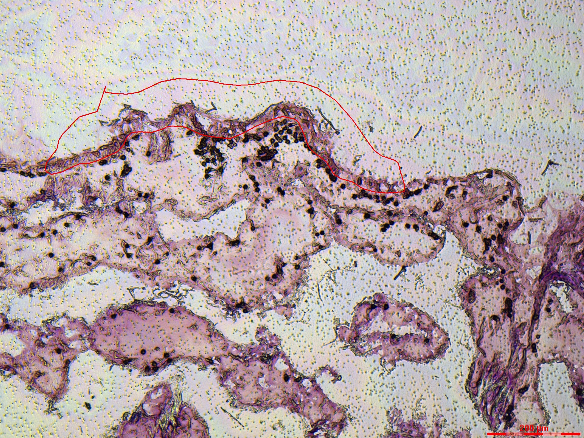
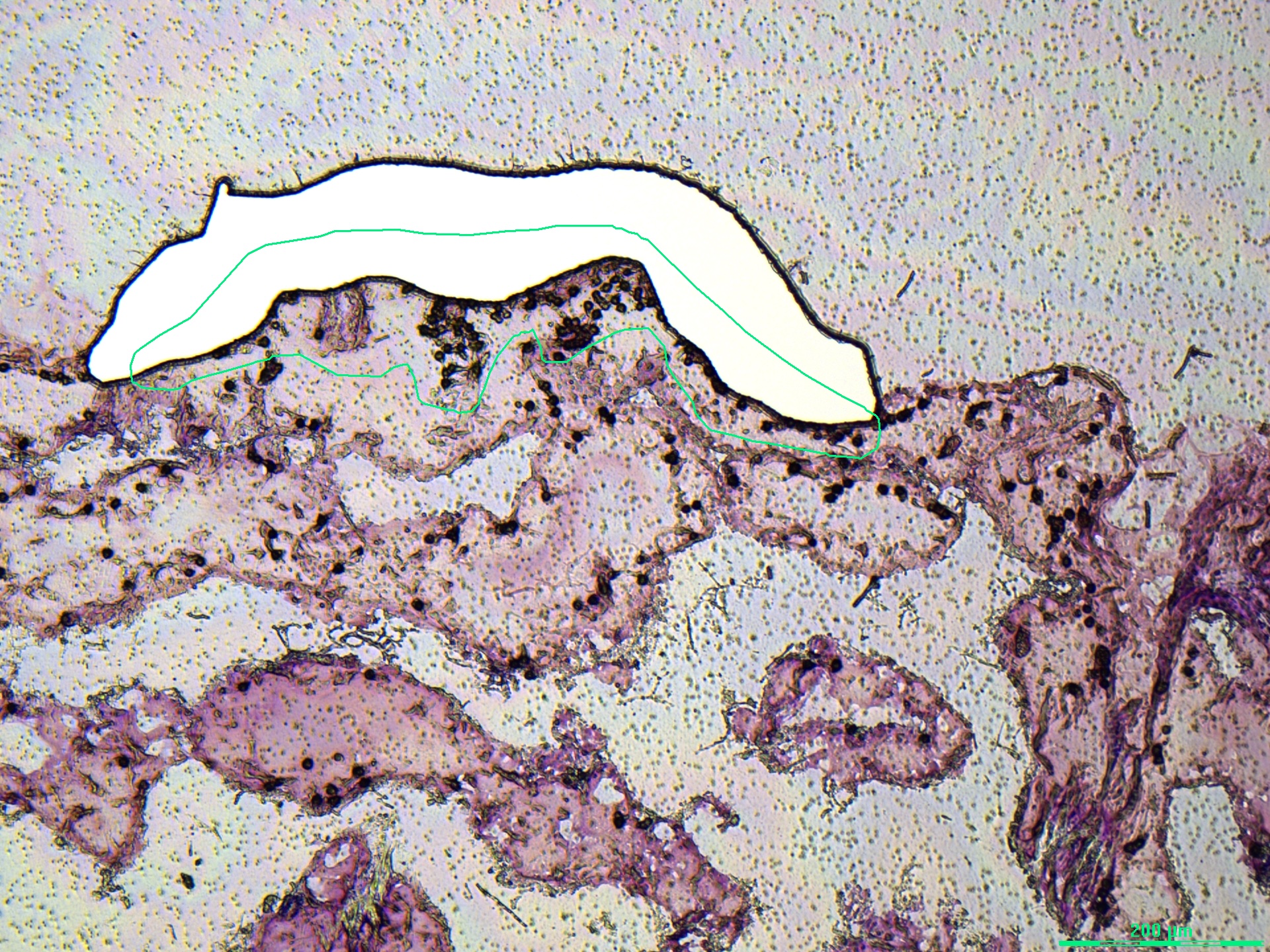
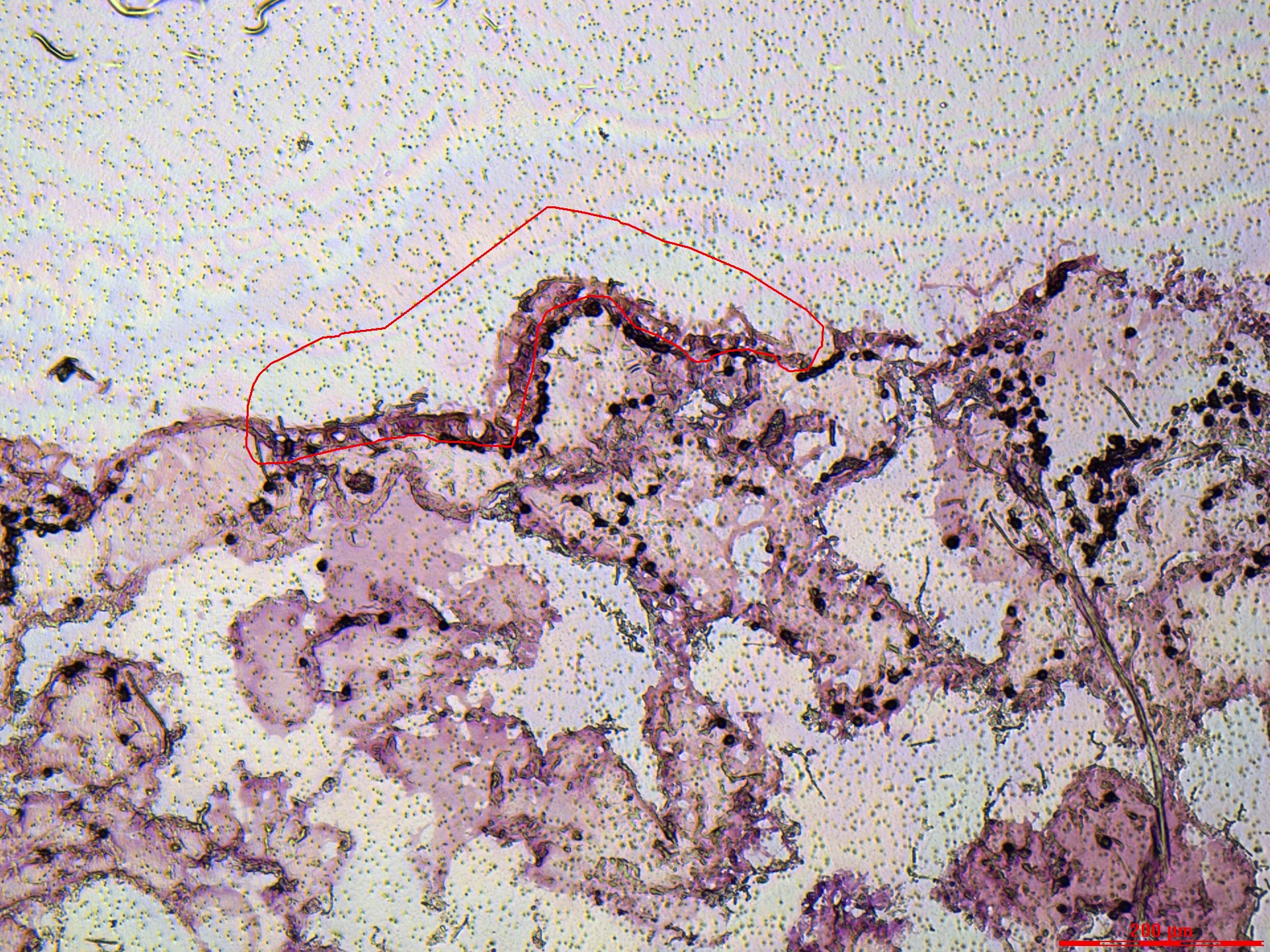
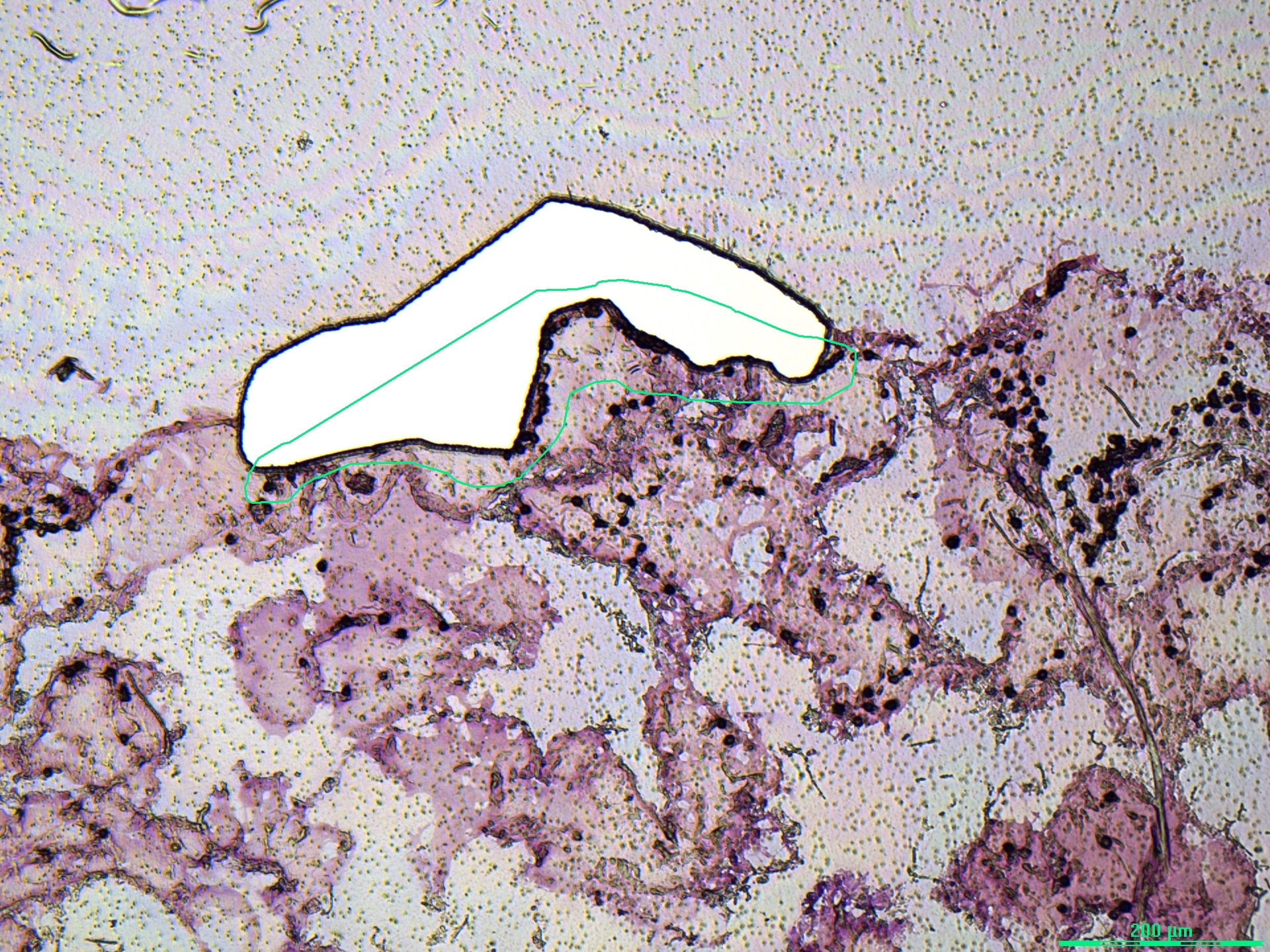
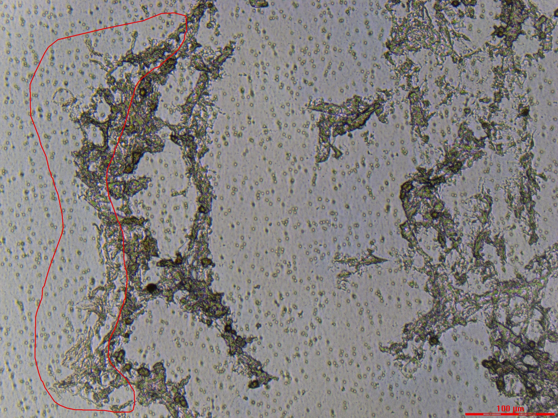
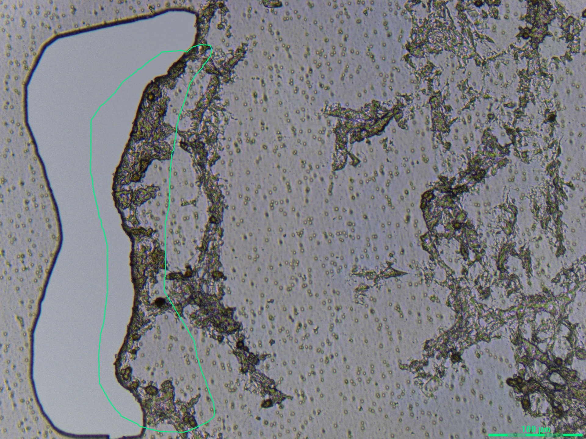
LCM Staining + Tissue Quality Assessment
Staining summary: Cresyl –> 70% EtOH –> RNase‑free water (brief) –> 100% EtOH
- Lots of purple stained OCT in tissue gaps, but tissue is overall looking okay for PEN membrane
- Pretty hard to distinguish oral epidermis and oral gastrodermis
- Slide 1 was better than Slide 2
Tubes collected: 1-13 (lost 6-8 in centrifuge…)
Data here: https://docs.google.com/spreadsheets/d/1iXE60arOpAE1sDYm0sR2PGPPH7mQKXzS5cIpavmqhCU/edit?usp=sharing
Example images:
Slide 1:
Slide 2:
| Oral Epidermis Dissection Attempt | Oral Gastrodermis Dissection Attempt |
|---|---|
 |
 |
 |
 |
Monday, 5/4/26
- Sectioned POR_4 from the 3/10/26 fixation onto 2 RNAse & UV-treated PEN membrane slides but changed one variable:
- Slide 1: Normal protocol (air dry –> 100% ethanol –> 15 min air dry in falcon tube –> dry ice)
- Slide 2: Normal protocol (no ethanol –> 2 min air dry in falcon tube –> dry ice)
Wednesday, 5/6/26 - Ethanol‑Only Variant (No RNase‑free Water; Added 75% Pre‑Step)
- Thawed and stained slides with modifications to the LCM Protocol
- Place slide on petri dish; on tube rack over dry ice (not immediately on the dry ice otherwise the 70% ethanol can freeze)
- Attempt to remove OCT by submerging ice-cold 75% ethanol for 2 mins
- Apply Cresyl violet staining solution (in 100% ethanol) directly with syringe and sterile filter to the section and incubate for 1 minute, swivel gently
- Incubated for closer to 30 seconds
- Rinse off stain by (pipetting) with ice-cold 70% ethanol
- And then place back on clean petri dish and cover slide with ice-cold 70% ethanol
- (ideally the ethanol just stays on the slide but if it rolls off then gently submerge the slide in 70% ethanol)
- Ideally this removes all the OCT. And hopefully no tissue.
- Be as gentle as possible and keep everything as cold as possible
If any OCT remains, rinse or gently submerge slide in ice-cold RNAse-free watermake sure this is not directly over dry ice but elevated, it will freeze if not
- Then submerge in or cover slide with ice-cold 100% ethanol for 1 minute to fully dry tissue and remove any excess water
- Air dry sample 1-2 in drying chamber with desiccant (or falcon tube with silica) or fume hood
- Proceed to LCM now, transport slide in falcon tube with silica packet
LCM Staining + Tissue Quality Assessment
Staining summary: 75% EtOH –> Cresyl –> 70% EtOH –> 100% EtOH
- TONS of OCT remained. Wasn’t stained super purple but there were sheets and folds of OCT that hampered dissections a lot
- Likely because of removing the RNAse free water step?
- The slide that had the ethanol step at the cryostat (Slide 1) looked marginally better than Slide 2 in tissue quality
- But no difference in OCT amounts
Tubes collected: 14-24
Data here: https://docs.google.com/spreadsheets/d/1iXE60arOpAE1sDYm0sR2PGPPH7mQKXzS5cIpavmqhCU/edit?usp=sharing
Example images:
Slide 1:
Slide 2:
| Oral Epidermis Dissection Attempt | Oral Gastrodermis Dissection Attempt |
|---|---|
 |
 |
 |
 |
 |
 |
Wednesday, 5/6/26 PM
- Sectioned one more slide POR_4 from the 3/10/26 fixation onto 1 RNAse & UV-treated PEN membrane slide and mimicked the slide 2 change from 5/4/26:
- Slide 3: No ethanol protocol (2 min air dry in falcon tube –> dry ice)
Monday, 5/11/26 - High‑Aqueous / PAXgene‑Like Protocol
- Thawed and stained slides with modifications to the LCM Protocol
- Increased aqueous protocol with 75%, 50%, 30% EtOH and multiple H2O steps (based on Paxgene protocol)
- 1 min 75% EtOH
- 45 s 50% EtOH
- 30 s 30% EtOH
- 30 s RNAse free H2O
- Cresyl violet, 1 minute
- Dip in 75% ethanol
- Another RNAse free H2O dunk to remove remaining OCT
- 100% ethanol 1 minute
- Increased aqueous protocol with 75%, 50%, 30% EtOH and multiple H2O steps (based on Paxgene protocol)
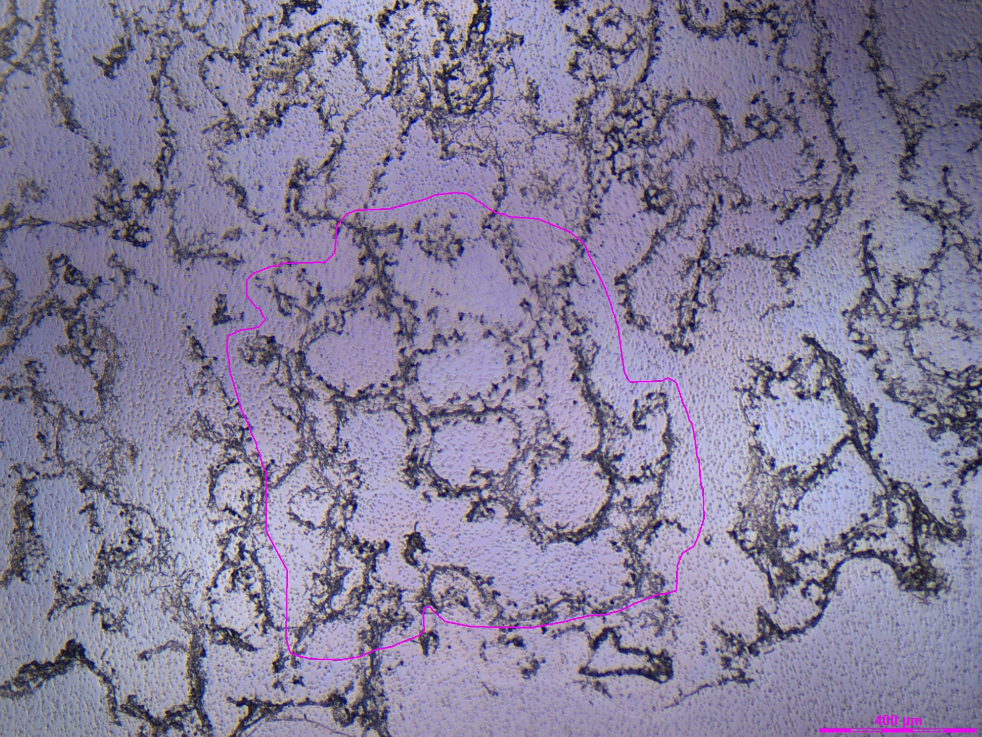
LCM Staining + Tissue Quality Assessment
Staining summary: 75% EtOH –> 50% EtOH –> 30% EtOH –> H2O –> Cresyl –> 75% EtOH –> H2O –> 100% EtOH
- Severe tissue loss. Too much water/washing.
- No ability to distinguish tissue layers
- When moving from the cold H2O to the cold 100% ethanol, i think the ethanol was SO cold that the water turned into ice in certain parts of the slide/tube, which looked not great
- Decided to still collect tissue for extraction testing, took big bulk pieces in different extraction buffers
- normal ProK digestion buffer from the LCM Protocol
- DNA/RNA shield
- RNA Lysis buffer from the extraction kit
- I also thawed Slide 2 from 4/28/26 which I didn’t collect any tissue from and did the same thing – collected cells in different buffers for testing (tubes 40-45)
Tubes collected: 31-39 & 40-45
Data here: https://docs.google.com/spreadsheets/d/1iXE60arOpAE1sDYm0sR2PGPPH7mQKXzS5cIpavmqhCU/edit?usp=sharing
Example images:
Monday, 5/11/26 PM - Ethanol-only staining attempt
- Sectioned from a new block (Time Series POR_R72_C2) onto 2 RNAse & UV-treated PEN membrane slide and mimicked the slide 2 change from 5/4/26:
- Slides 1&2: No ethanol protocol (2 min air dry in falcon tube –> dry ice)
- Stained Slide 1 right away
- 3 min 70% EtOH
- 2 min 70% EtOH
- 30s 96% EtOH
- 30s 100% EtOH
- Cresyl violet 1 minute
- 1 min 70% EtOH
- Extra rinse with 70% by pipetting to try to remove OCT
- 30s 96% EtOH
- 100% EtOH 1 minute
- Dried in fume hood
LCM Staining + Tissue Quality Assessment (5/13/26)
Staining summary: Cryostat (no overnight freeze) –> 70% EtOH –> 96% EtOH –> 100% EtOH –> Cresyl –> 70% EtOH –> 96% EtOH –> 100% EtOH
- Went to the scope on 5/11 but did not collect any samples. Collected samples on 5/13
- There was so much OCT, worse than 4/28
- Very purple and hard to distinguish tissues, but did some tests
- Although there appeared to be a lot of OCT and very dark stain that made seeing the tissues hard, the dissection/laser cutting was surprisingly easy compared to 5/6/26 for example
Tubes collected on 5/13/26: 60-63
| Oral Epidermis Dissection Attempt | Oral Gastrodermis Dissection Attempt |
|---|---|
 |
 |
 |
 |
Wednesday, 5/13/26
- Thawed POR_R72_C2 Slide 2 and did wash steps to remove OCT only, no staining (all in petri dish over dry ice, all solutions ice cold)
- 3 min 70% EtOH
- 2 min 70% EtOH
- 10 s RNAse free H2O
- 30s 70% EtOH
- 30s 96% EtOH
- 1 min 100% EtOH
- Dried in fume hood
LCM Staining + Tissue Quality Assessment
Staining summary: No staining
- Honestly looks okay without the stain, but the stain helps a little bit to distinguish tissues and removing it clearly still didn’t solve all my problems
- There was still tissue loss, likely from the RNAse free water step
Tubes collected: 51-58
Data here: https://docs.google.com/spreadsheets/d/1iXE60arOpAE1sDYm0sR2PGPPH7mQKXzS5cIpavmqhCU/edit?usp=sharing
Example images:
| Oral Epidermis Dissection Attempt | Oral Gastrodermis Dissection Attempt |
|---|---|
 |
 |
 |
 |
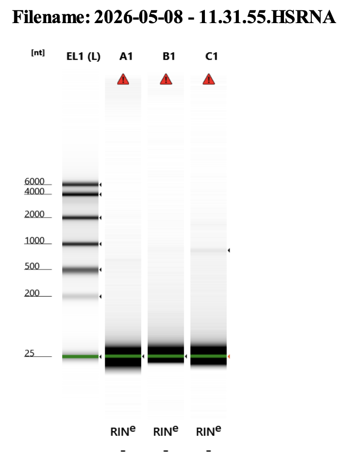
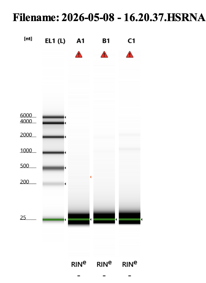
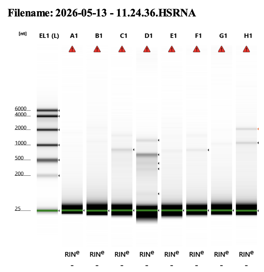
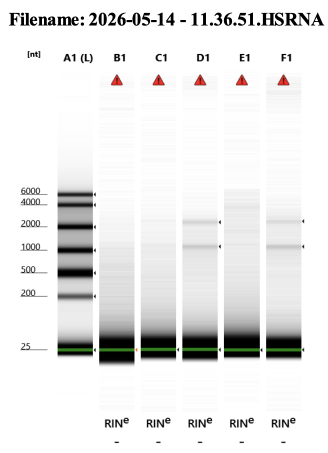
RNA extraction results (up to 5/14/26):
My RNA concentrations are much much lower than I am used to for LCM.
Extraction Protocol Here. I follow this closely, but there are some modifications I have tried in increase yield, noted in the table below:
- Different cell collection buffers (my normal PK buffer, DNA/RNA shield, or RNA Lysis buffer)
- Different digestion times
- 15 min RT = 15 minutes room temperature
- 15 min 56C 1400rpm = 15 minutes at 56 ºC on thermomixer 1400 rpm
- 1 hr 56C 1400rpm = 1 hour at 56 ºC on thermomixer 1400 rpm
- 1 hr 52C = 1 hour at 52 ºC in the Incubator Genie, gentle shaking
- Overnight 56C = Overnight (5pm-10:30am) at 56 ºC in the Incubator Genie, no shaking
Best results seem to come from 1 hr 56C 1400rpm protocol.
| Slide | Section | Tube # | Number dissections | Tissue | Extraction Buffer | Notes | Total Dissection Area | Digestion | Tapestation assessment 1-3 | TS Conc (pg/uL) | pg in 8 uL | pg/area |
|---|---|---|---|---|---|---|---|---|---|---|---|---|
| POR_3_Slide1 | 5 | 1 | 5 | oral epidermis | PK Buffer | extracted 5/13/26 | 160,516 | 15 min RT | 1 | 44.6 | 357 | 0.0022 |
| POR_3_Slide1 | 5 | 2 | 5 | oral gastrodermis | PK Buffer | extracted 5/13/26 | 86,158 | 1 hr 56C 1400rpm | 1 | 54.9 | 439 | 0.0051 |
| POR_3_Slide1 | 4 | 9 | 1 | bulk - all tissues | PK Buffer | extracted 5/13/26 | 3,215,616 | 15 min RT | 2 | 48.1 | 385 | 0.0001 |
| POR_3_Slide1 | 1 | 13 | 2 | bulk - all tissues | PK Buffer | extracted 5/13/26 | 6,681,317 | 1 hr 56C 1400rpm | 3 | 108.0 | 864 | 0.0001 |
| POR_4_Slide1 | 1 | 17 | 3 | oral epidermis | PK Buffer | extracted 5/8/26 PM | 234,535 | 1 hr 52C | 1 | 33.0 | 264 | 0.0011 |
| POR_4_Slide1 | 1 | 18 | 3 | oral gastrodermis | PK Buffer | extracted 5/8/26 PM | 183,347 | 1 hr 52C | 1 | 33.9 | 271 | 0.0015 |
| POR_4_Slide1 | 4 | 21 | 5 | oral epidermis | PK Buffer | extracted 5/8/26 AM | 346,744 | 15 min RT | 1 | 43.5 | 348 | 0.0010 |
| POR_4_Slide1 | 4 | 22 | 6 | oral gastrodermis | PK Buffer | extracted 5/8/26 AM | 303,230 | 15 min RT | 1 | 44.7 | 358 | 0.0012 |
| POR_4_Slide1 | 5 | 23 | 1 | bulk - all tissues | PK Buffer | extracted 5/8/26 PM | 830,657 | 1 hr 52C | 2 | 33.1 | 265 | 0.0003 |
| POR_4_Slide1 | 5 | 24 | 1 | bulk - all tissues | PK Buffer | extracted 5/8/26 AM | 1,002,718 | 15 min RT | 2 | 38.3 | 306 | 0.0003 |
| POR_4_Slide3 | 1 | 31 | 1 | bulk | DNA/RNA Shield | extracted 5/13/26 | 2,401,591 | 15 min RT | 1 | 46.3 | 370 | 0.0002 |
| POR_4_Slide3 | 2 | 32 | 1 | bulk | DNA/RNA Shield | extracted 5/13/26 | 2,575,547 | 15 min 56C 1400rpm | 2 | 46.1 | 369 | 0.0001 |
| POR_4_Slide3 | 4 | 33 | 1 | bulk | RNA Lysis Buffer | extracted 5/13/26 | 1,861,138 | 15 min RT | 1 | 48.4 | 387 | 0.0002 |
| POR_4_Slide3 | 5 | 34 | 1 | bulk | PK Buffer | extracted 5/13/26 | 2,534,590 | 15 min 56C 1400rpm | 3 | 65.9 | 527 | 0.0002 |
| POR_R72_C2_2_unstained | 2 | 51 | 5 | oral epidermis | PK Buffer | extracted 5/14/26 | 110,765 | Overnight 56C | 1 | 31.4 | 251 | 0.0023 |
| POR_R72_C2_2_unstained | 2 | 52 | 5 | oral gastrodermis | PK Buffer | extracted 5/14/26 | 84,931 | 1 hr 56C 1400rpm | 1 | 27.1 | 217 | 0.0026 |
| POR_R72_C2_2_unstained | 3 | 55 | 6 | oral epidermis | PK Buffer | extracted 5/14/26 | 276,815 | 1 hr 56C 1400rpm | 2 | 24.4 | 195 | 0.0007 |
| POR_R72_C2_1 | 2 | 60 | 5 | oral epidermis | PK Buffer | extracted 5/14/26 | 343,268 | 1 hr 56C 1400rpm | 1 | 35.4 | 283 | 0.0008 |
| POR_R72_C2_1 | 2 | 61 | 5 | oral gastrodermis | PK Buffer | extracted 5/14/26 | 239,654 | 15 min 56C 1400rpm | 2 | 26.5 | 212 | 0.0009 |
 |
17, 18, 21 |
 |
22, 23, 24 |
 |
1, 2, 9, 13, 31, 32, 33, 34 |
 |
51, 52, 55, 60, 61 |
RNA Library Prep:
As noted above, my RNA concentrations are much much lower than I am used to for LCM. But I still want to test these samples to see what we can get. So started library prep on 5/14/26 for the following samples:
| Slide | Section | Tube # | Number dissections | Tissue | Extraction Buffer | Notes | Total Dissection Area | Digestion | Tapestation assessment 1-3 | TS Conc (pg/uL) | pg in 8 uL | pg/area |
|---|---|---|---|---|---|---|---|---|---|---|---|---|
| POR_3_Slide1 | 1 | 13 | 2 | bulk - all tissues | PK Buffer | extracted 5/13/26 | 6,681,317 | 1 hr 56C 1400rpm | 3 | 108.0 | 864 | 0.0001 |
| POR_4_Slide3 | 5 | 34 | 1 | bulk | PK Buffer | extracted 5/13/26 | 2,534,590 | 15 min 56C 1400rpm | 3 | 65.9 | 527 | 0.0002 |
| POR_R72_C2_1 | 2 | 61 | 5 | oral gastrodermis | PK Buffer | extracted 5/14/26 | 239,654 | 15 min 56C 1400rpm | 2.0 | 26.5 | 212 | 0.0009 |
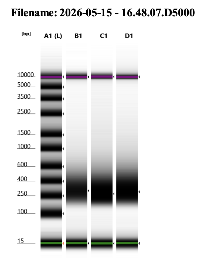
5/15/26 cDNA Results
I used 20 cycles for cDNA amplification (Step 2.4.3). They look pretty great!
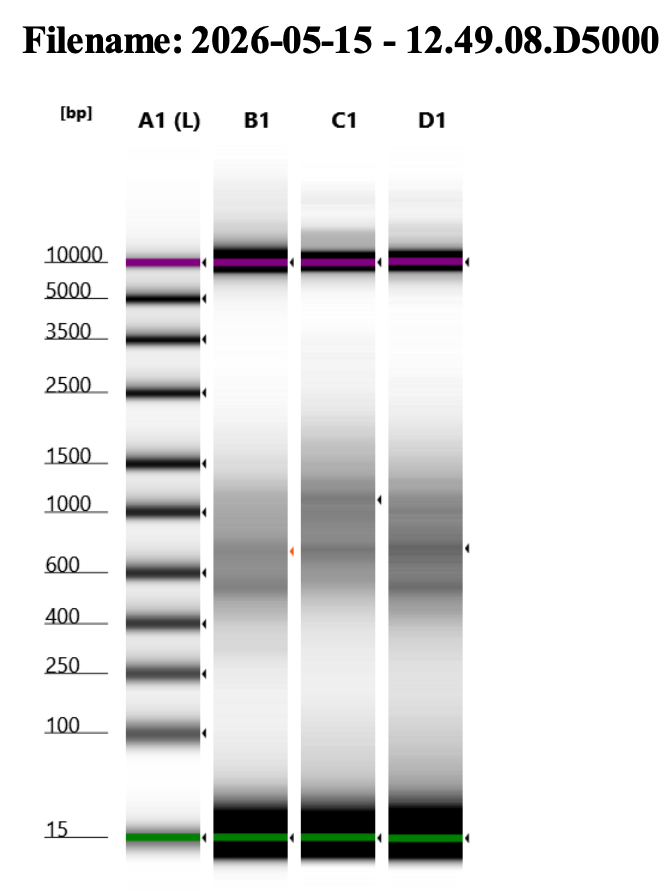
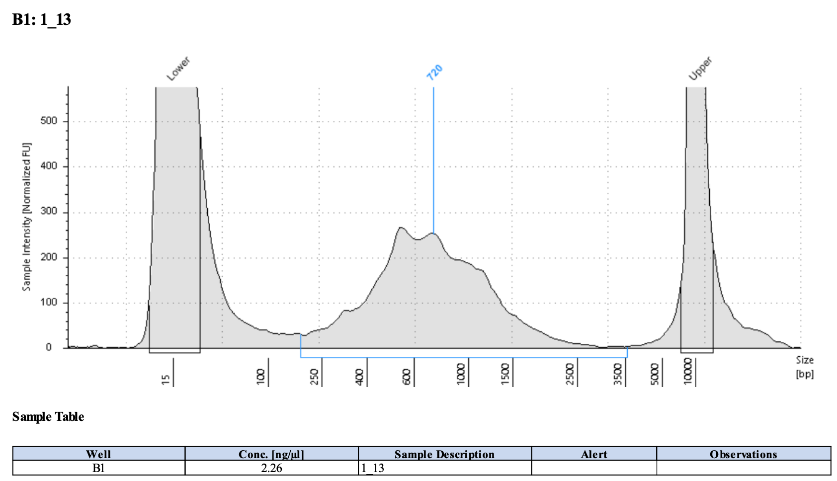
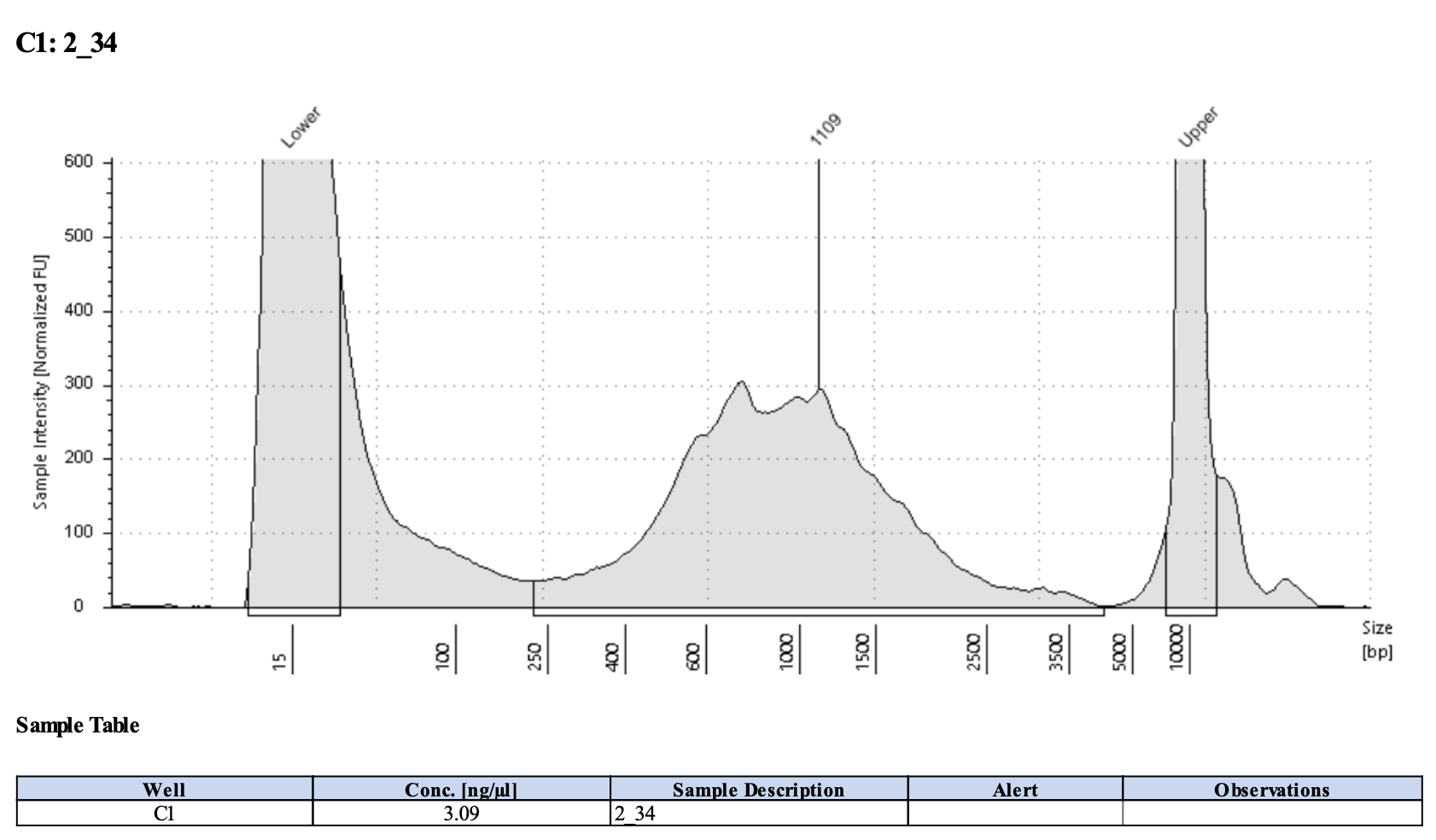
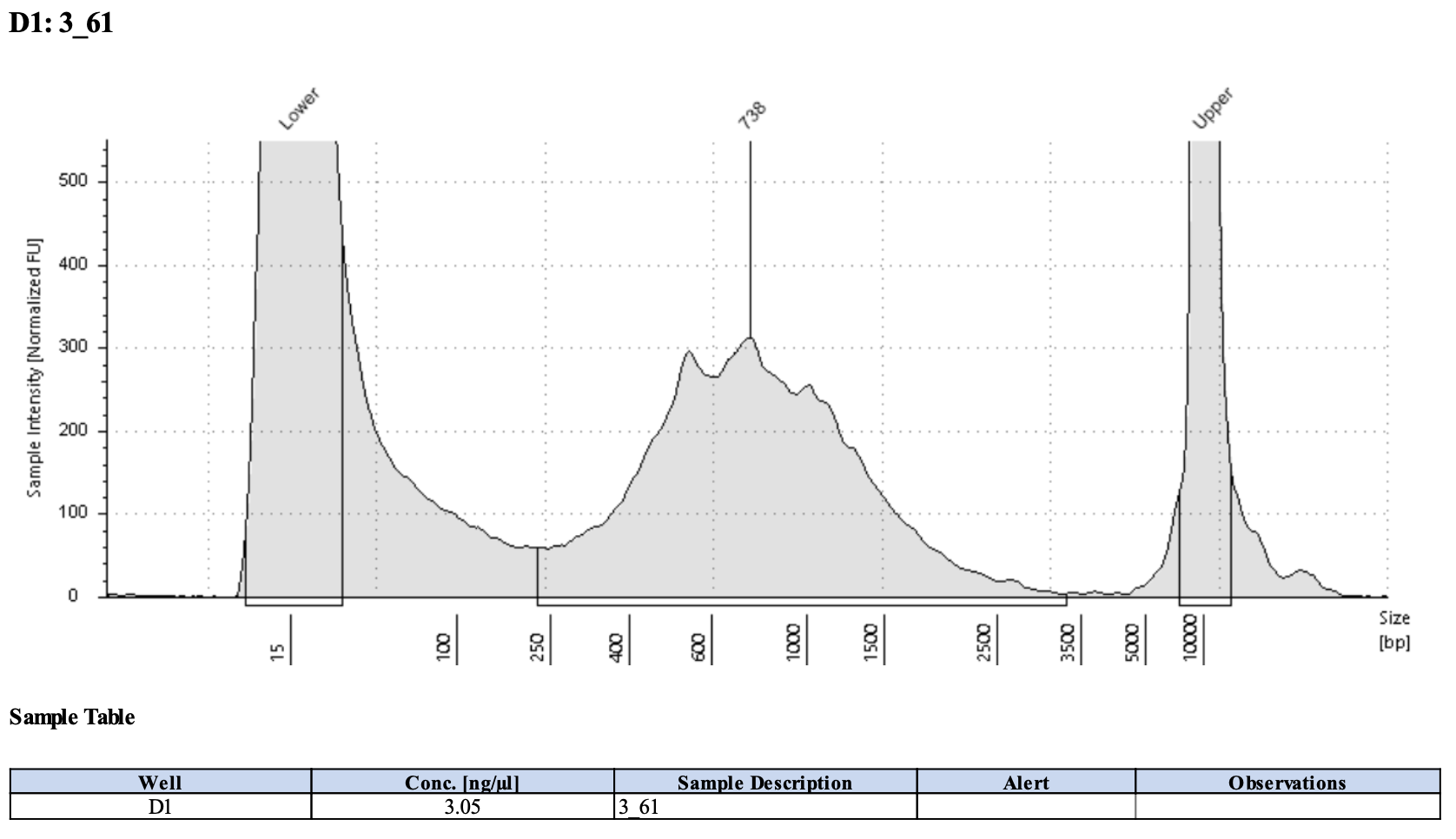
cDNA and planned cycles/indeces
| Sample Type | cDNA concentration | amount cDNA input (conc * 26 uL) | PCR cycles for final step | Index_ID | index |
|---|---|---|---|---|---|
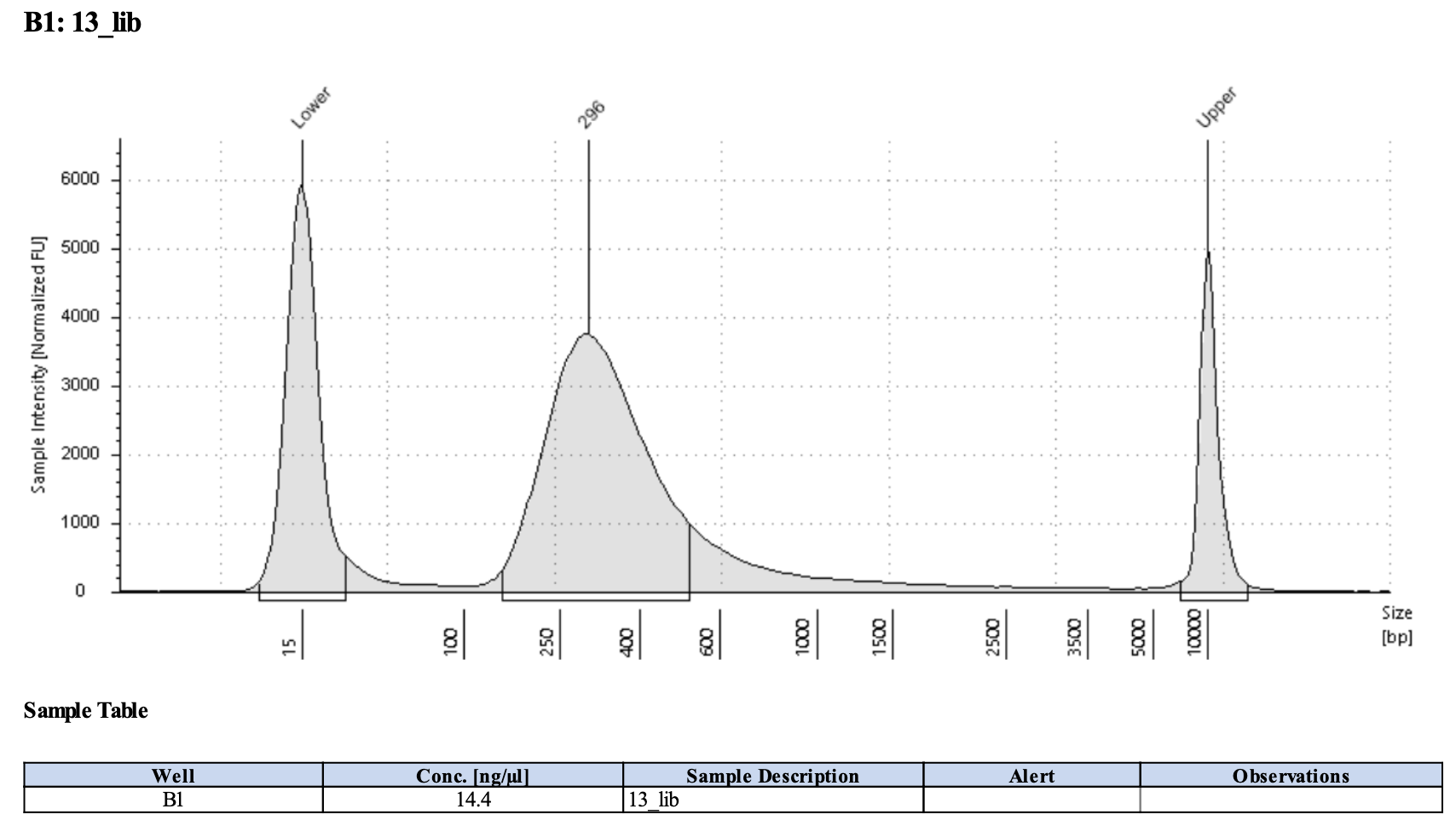
| #13 | 2.26 | 58.76 | E7500S-18 | GTCCGC | |
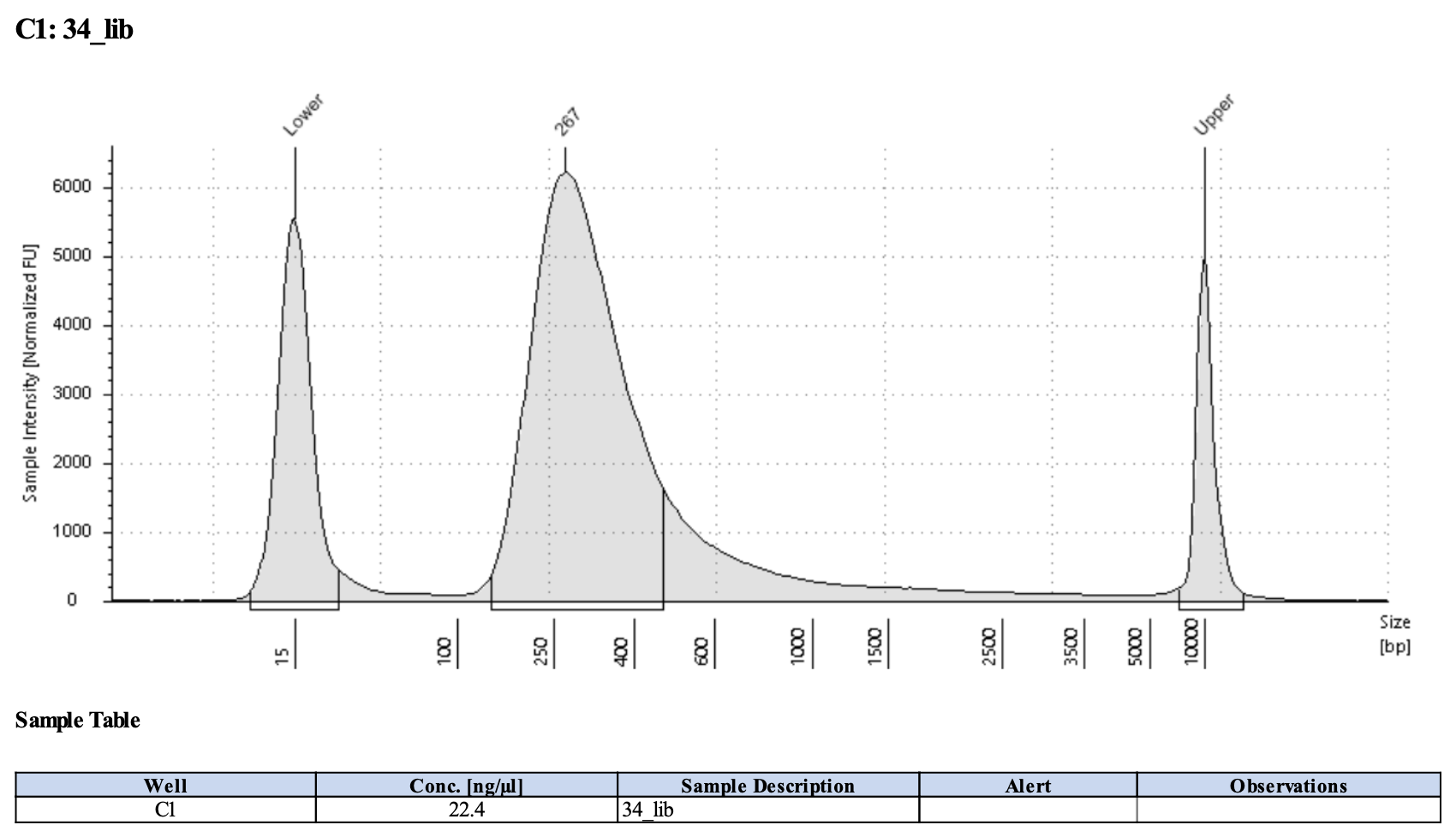
| #34 | 3.09 | 80.34 | E7500S-22 | CGTACG | |
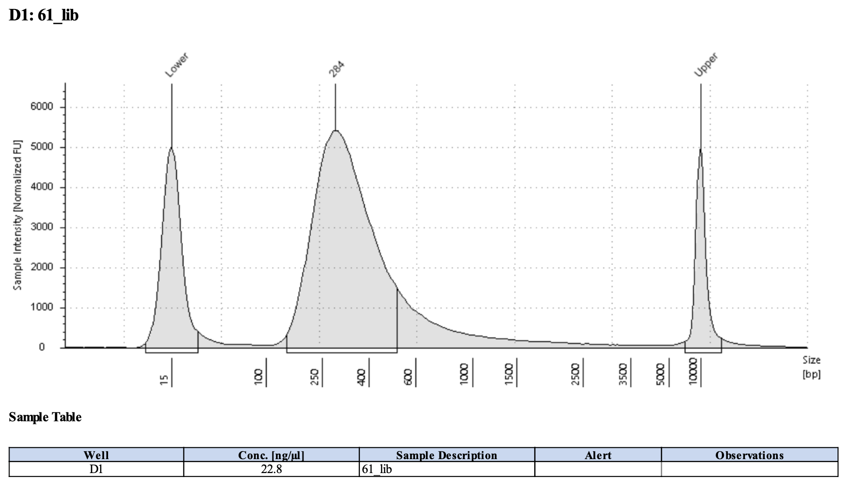
| #61 | 3.05 | 79.30 | E7500S-23 | GAGTGG |
Full results can be found here.
5/15/26 Final Library Results
I used 8 cycles for Final Library amplification (Step 2.10.3). They look amazing!!
Full results can be found here.
Pooling
Submitting a pool of 50 uL at 4 nM concentration.
Current thoughts
- Original protocol of 70% EtOH + RNase‑free water + 100% EtOH gives the best OCT removal with tissue present but tissue quality still isn’t ideal
- Ethanol‑only (75/96/100) is safer for tissue adhesion but leaves too much OCT to see tissue layers
- PAXgene‑like (75/50/30 + water) protocol is really good for OCT removal but the tissue is horrendous
Further testing
Thursday, 5/21/26
Sectioned two slides each of POR_R3_H1 & POR_R72_H1. Did 12 um thick sections. Dried 2 mins in cryostat, then put in 50 mL tube with silica packet at room temperature for 15 minutes, then transferred to dry ice.
Friday AM, 5/22/26
- Cover with ice-cold 70% ethanol for 2-3 mins
- Replace with fresh ice-cold 70% ethanol for 2 mins
- Replace with 100% ethanol for 30s
- Apply cresyl violet staining (in 100% ethanol), incubate 30 seconds
- Pour off excess stain
- Cover slide with ice-cold 70% ethanol, 30s
- Pour off excess stain
- Cover slide with fresh ice-cold 70% ethanol, 30s
- If any OCT remains, rinse or gently submerge slide in ice-cold RNAse-free water for 10s
- Then immediately back into ice-cold 70% ethanol
- Dehydrate:
- 30-60s 70% EtOH
- 30s 96% EtOH
- 1 min 100% EtOH
- Dry in fume hood
LCM Staining + Tissue Quality Assessment
Did not look good. Collected tubes 101-108. Tube 107 spilled.
Friday PM, 5/22/26
- Did all staining in reagent troughs
- Cover with ice-cold 70% ethanol for 2-3 mins
- Dunk in cold RNAse free water 5-6 times to try to remove OCT
- Dunk in 70% ethanol to remove water
- Apply cresyl violet staining (in 100% ethanol), incubate 30 seconds
- Dunk in 70% ethanol to remove stain
- Cover with ice-cold 70% ethanol for 30s
- Cover slide with 100% ethanol, 1 minute
- Dry in fume hood
LCM Staining + Tissue Quality Assessment
POR R72 H1 slide was basically ruined, all the tissue came off while dunking in water. POR R3 H1 worked okay but looked bad on the scope. A lot of tissue loss.
Collected 2 tubes 109-110.
Tuesday, 5/26/26
- POC R3 C2 - 2 slides, one with 70% ethanol at cryostat one without
- POR R72 H1 - 1 slide with 70% ethanol at cryostat
- Ones with ethanol:
- Dried 1 minute in cryostat
- Dried 1 minute at RT in petri dish
- Covered with cold 70% ethanol for 30s-1 min with swirling
- Dried and then put in falcon tube with silica to dry fully at RT for 10-15 mins before putting on dry ice
Wednesday AM, 5/27/26
Try standard protocol on the POC slides.
Procedure:
- Morning of LCM: Bring slide up to room temperature, slowly to avoid formation of water condensation inside the container. Did the following:
- 30 minutes at -20 ºC
- 30 minutes at 4°C
- 15 minutes at room temp
- Place slide on petri dish; on tube rack over dry ice (not immediately on the dry ice otherwise the 70% ethanol can freeze)
- Apply Cresyl violet staining solution (in 100% ethanol) directly with syringe and sterile filter to the section and incubate for 1 minute, swivel gently
- Rinse off stain by (pipetting) with ice-cold 70% ethanol
- And then place back on clean petri dish and cover slide with ice-cold 70% ethanol
- (ideally the ethanol just stays on the slide but if it rolls off then gently submerge the slide in 70% ethanol)
- Ideally this removes all the OCT. And hopefully no tissue.
- Be as gentle as possible and keep everything as cold as possible
- If any OCT remains, rinse or gently submerge slide in ice-cold RNAse-free water
- make sure this is not directly over dry ice but elevated, it will freeze if not
- Then submerge in or cover slide with ice-cold 100% ethanol for 1 minute to fully dry tissue and remove any excess water
- Air dry sample 1-2 in drying chamber with desiccant (or falcon tube with silica) or fume hood
- Proceed to LCM now, transport slide in falcon tube with silica packet
LCM Staining + Tissue Quality Assessment
POC slides looked good, the one without 70% ethanol was marginally better than the one with. However, I want to keep in mind that my original POC success ahd a 100% ethanol step at the cryostat, which I should consider again.
Collected tubes 201-214.
Extraction notes: didn’t go super well. Really low concentrations for POC.
Friday, 5/29/26
- POR R72 C2 - 2 slides with 100% ethanol at cryostat
- Dried 1 minute in cryostat
- Dried 1 minute at RT in petri dish
- Covered with cold 100% ethanol for 30s-1 min with swirling
- Dried and then put in falcon tube with silica to dry fully at RT for 10-15 mins before putting on dry ice
Procedure:
- Same day LCM: Bring slide up to room temperature, slowly to avoid formation of water condensation inside the container. Did the following:
- 30 minutes at -20 ºC
- 30 minutes at 4°C
- 15 minutes at room temp
- Place slide on petri dish; on tube rack over dry ice (not immediately on the dry ice otherwise the 70% ethanol can freeze)
SLIDE 1 (Pre-wash):
- Cover with ice-cold 70% ethanol to remove excess OCT, 30s
- Replace with fresh ice-cold 70% ethanol
- Cover with ice-cold 96% ethanol, 10 s
- Apply Cresyl violet staining solution (in 100% ethanol) directly with syringe and sterile filter to the section and incubate for 30s, swivel gently
- Rinse off stain by (pipetting) with ice-cold 100% ethanol
- Will need to rinse a few times to remove all the stain
- Then submerge in or cover slide with ice-cold 100% ethanol for 1 minute to fully dry tissue and remove any excess water
- Air dry sample 1-2 in drying chamber with desiccant (or falcon tube with silica) or fume hood
- Proceed to LCM now, transport slide in falcon tube with silica packet
SLIDE 2:
- Apply Cresyl violet staining solution (in 100% ethanol) directly with syringe and sterile filter to the section and incubate for 30s, swivel gently
- Rinse off stain by (pipetting) with ice-cold 100% ethanol
- And then place back on clean petri dish and cover slide with ice-cold 70% ethanol to remove excess stain and OCT
- Will need to rinse a few times to remove all the stain
- Cover with ice-cold 96% ethanol, 10 s
- Then submerge in or cover slide with ice-cold 100% ethanol for 1 minute to fully dry tissue and remove any excess water
- Air dry sample 1-2 in drying chamber with desiccant (or falcon tube with silica) or fume hood
- Proceed to LCM now, transport slide in falcon tube with silica packet
LCM Staining + Tissue Quality Assessment
The prewashed slide was easier to cut but outside tissue layers looked less intact than the non-prewashed slide. But the non-prewashed slide had a ton of OCT left and a lot of cracking of the OCT. Difficult to cut due to so much OCT.
Collected tubes 215-224.
| Slide | Section (#d from labelled part of slide) | Tube # | Number dissections | Tissue | Extraction Buffer | Notes | Total Dissection Area | Total Dissection Area / 40 (approx # cells) | Approx Tissue Area, quick measurements – will redo | Ratio Dissection/Tissue | Approx Tissue Area / 40 (approx # cells) |
|---|---|---|---|---|---|---|---|---|---|---|---|
| POR_R72_C2_2_529 | 3 | 215 | 6 | oral epidermis | PK Buffer | extracted 5/30 | 405,830 | 10,146 | 113,703 | 3.57 | 2843 |
| POR_R72_C2_2_529 | 3 | 216 | 7 | oral gastrodermis | PK Buffer | extracted 5/30 | 334,040 | 8,351 | 165,866 | 2.01 | 4147 |
| POR_R72_C2_2_529 | 2 | 217 | 3 | oral epidermis | PK Buffer | 213,808 | 5,345 | 45,312 | 4.72 | 1133 | |
| POR_R72_C2_2_529 | 2 | 218 | 3 | oral gastrodermis | PK Buffer | 160,971 | 4,024 | 86,392 | 1.86 | 2160 | |
| POR_R72_C2_2_529 | 4 | 219 | 2 | bulk | PK Buffer | extracted 5/30 | 442,544 | 11,064 | 293,360 | 1.51 | 7334 |
| POR_R72_C2_2_529 | 5 | 220 | 3 | bulk | PK Buffer | 425,766 | 10,644 | 311,347 | 1.37 | 7784 | |
| POR_R72_C2_1_529_Prewash | 1 | 221 | 2 | oral epidermis | PK Buffer | tissue looked worse but way easier to cut | 177,470 | 4,437 | 29,699 | 5.98 | 742 |
| POR_R72_C2_1_529_Prewash | 1 | 222 | 2 | oral gastrodermis | PK Buffer | 85,555 | 2,139 | 53,941 | 1.59 | 1349 | |
| POR_R72_C2_1_529_Prewash | 1 | 223 | 1 | bulk | PK Buffer | 1,325,972 | 33,149 | 828,061 | 1.60 | 20702 | |
| POR_R72_C2_1_529_Prewash | 4 | 224 | 1 | bulk | PK Buffer | 1,063,735 | 26,593 | 581,598 | 1.83 | 14540 |
Extraction: 5/30/26
| Slide | Section (#d from labelled part of slide) | Tube # | Number dissections | Tissue | Extraction Buffer | Notes | Total Dissection Area | Digestion | Tapestation assessment worst (1)-best(3) | TS Conc (pg/uL) | pg in 8 uL | pg/area |
|---|---|---|---|---|---|---|---|---|---|---|---|---|
| POR_R72_C2_2_529 | 2 | 216 | 5 | oral gastrodermis | PK Buffer | extracted 5/30/26 | 334,040 | 15 min 56C 1400rpm | 2 | 60.3 | 482 | 0.0014 |
| POR_R72_C2_2_529 | 2 | 215 | 5 | oral epidermis | PK Buffer | extracted 5/30/26 | 405,830 | 15 min 56C 1400rpm | 2 | 42.7 | 342 | 0.0008 |
| POR_R72_C2_2_529 | 2 | 219 | 5 | bulk | PK Buffer | extracted 5/30/26 | 442,544 | 15 min 56C 1400rpm | 1 | 35.0 | 280 | 0.0006 |
Extraction validation: Is the issue the kit or just the volume of dissected tissue?
“Bulk” below refers to fresh adult tissue in DNA/RNA shield tube. Bulk in all instances above refers to microdissected tissues that were not tissue-layer specific but instead were big chunks of tissue.
| Tube | Sample Type | Input / Source | PK Digestion | Kit | Elution (µL) | Conc (pg/µL) | Total RNA (pg) | Total RNA (ng) | dv200 (% >200 nt) | Notes |
|---|---|---|---|---|---|---|---|---|---|---|
| 1A | Fresh tissue in DNA/RNA Shield | 150 µL Shield | 15 min RT | MicroPrep | 15 | 3,770 | 56,550 | 56.6 | 81.6 | Good |
| 1B | Fresh tissue in DNA/RNA Shield | 150 µL Shield | 15 min 56 °C, 1400 rpm | MicroPrep | 15 | 4,740 | 71,100 | 71.1 | 81.0 | More degraded |
| 1C | Fresh tissue in DNA/RNA Shield | 150 µL Shield | 1 hr 56 °C, 1400 rpm | MicroPrep | 15 | 4,460 | 66,900 | 66.9 | 55.0 | Over‑digested, really degraded |
| 1D | Fresh tissue in DNA/RNA Shield (control) | 300 µL Shield | 15 min RT | Miniprep | 100 | 5,270 | 527,000 | 527 | 79.7 | Best extraction, followed my standard DNA/RNA shield bulk tissue protocol |
| Tube | Sample Type | Source | PK Digestion | Kit | Elution (µL) | Conc (pg/µL) | Total RNA (pg) | Total RNA (ng) | dv200 (% >200 nt) | Notes |
|---|---|---|---|---|---|---|---|---|---|---|
| 2A | Whole Porites section, prewashed slide | 1 section from POR_R72_C2_1_Prewash | 15 min RT | MicroPrep | 15 | 157 | 2,355 | 2.36 | 70.2 | Very good |
| 2B | Whole Porites section, prewashed slide | 1 section from POR_R72_C2_1_Prewash | 15 min 56 °C, 1400 rpm | MicroPrep | 15 | 191 | 2,865 | 2.87 | 68.3 | More degraded than 2A |
| 2C | Whole Porites section, prewashed slide | 1 section from POR_R72_C2_1_Prewash | 1 hr 56 °C, 1400 rpm | MicroPrep | 15 | 133 | 1,995 | 2.00 | 57.3 | Still >50% dv200, more smear-y |